

Without physical optics there would have been no microscope, and until the perfection of the microscope, the biologist was in the main limited to what his unaided senses told him. It was by means of the compound microscope that the development of the cell theory was made possible in the nineteenth century. Similarly it is by means of the new tools and techniques developed in many instances by the physical sciences that the door to a biology of molecules has only recently been opened.
Warren Weaver, Rockefeller Foundation, and Molecular Biology
From 1931 to 1959, Warren Weaver ran the Rockefeller Foundation’s (RF) Natural Sciences Division. A mathematician with no background in biology, Weaver did something unusual for a philanthropy: he took roughly 80% of his division’s budget and concentrated it on a single thesis — funding the application of physics techniques to biological problems.
His maverick approach to science philanthropy, strong conviction, and interdisciplinary transgression paid off. Nearly every Nobel Prize in molecular biology awarded between 1954 and 1965 traces back to a Rockefeller Foundation grant — grants made, on average, two decades before the prize. It remains the single clearest case study of how concentrated, high-conviction science funding can generate a burst of new discoveries and ultimately create an entirely new scientific field, which Weaver dubbed “molecular biology”.1
Warren Weaver and New Science Instruments
In the two decades since [the Rockefeller Foundation made the decision to concentrate funding in molecular biology], there has been a remarkable development in the methods of biological investigation. The change is especially marked in the application of physical tools and techniques, such as the ultracentrifuge, the electrophoresis apparatus, spectroscopy, X-ray diffraction, the electron microscope and isotopic tracers. Through grants and fellowships the Foundation contributed to the development of some of these tools of research and to the application and extension of all of them to biological problems.
How did Warren Weaver fund and create the field of molecular biology? Others have written about how he brought in physicists to tackle biology problems, or the unique “fox”-shaped project officers Weaver recruited to identify and fund scientists, but I want to add a new dimension to Weaver’s strategy to creating a new field of science.
In “A Quarter Century in the Natural Sciences”, an essay Weaver wrote as a reflection on his time at Rockefeller Foundation, he is quite explicit that the field of molecular biology was created through new science instruments. Specifically, through:
New methods of separation: ultracentrifuge and electrophoresis
New methods of seeing: electron microscope, x-ray crystallography, and radioactive isotope tagging
Why are new science instruments so critical to creating new science fields? Because new science instruments create new ways of perceiving the world. Nearly every one of the Nobel Prizes that Weaver would go on to fund can be attributed to one of the 5 instruments/techniques listed above.
How did Weaver incorporate new science instruments into his field creation strategy? Analyzing Rockefeller Foundation annual reports from 1930-1950 and secondary sources, I will use the ultracentrifuge, electron microscope, and x-ray crystallography as case studies of how Weaver drove the application of these new instruments to the problems of biology, identify specific patterns of how Weaver drove the development and adoption of these new science instruments to ultimately lead to Nobel Prize winning discoveries, and what we can learn from Weaver’s use of new science instruments to drive field creation.
Ultracentrifuge
I too have made a wonderful journey…For the last 25 years, I have roamed through living cells, but with the help of a centrifuge rather than of a microscope.
While centrifugation had long been used to separate substances of different densities, the ultracentrifuge generated centrifugal forces orders of magnitude greater, allowing separation of even different proteins, and crucially integrated real-time optical detection to measure the properties of separated components. Theodor Svedberg developed the first ultracentrifuge to better understand colloidal chemistry, which won him the Nobel Prize in 1926, both for his experimental results and the development of a new science instrument that enabled those findings, one of the few Nobels awarded for instrumentation.
But adoption was slow, as Svedberg’s ultracentrifuge was bulky and quite dangerous — massive rotational forces meant if it came apart, and early versions often did, it would do so quite violently. In 1931, Weaver funded Stanford professor James McBain to spend a year with Svedberg, to learn about the ultracentrifuge. McBain would go on to build several simpler ultracentrifuges for use in the U.S. In 1934, Weaver funded Professor Beams’ team at UVA to develop a new, electrically driven ultracentrifuge, rather than oil-turbine design of Svedberg. It was this electrical ultracentrifuge design that was ultimately commercialized at scale.
The early days of new science instruments are often quite hacky, with tinkerer scientists engineering their own contraptions. Weaver’s support of multiple independent efforts to build ultracentrifuges helped drive down costs and accelerate eventual commercial deployment of ultracentrifuges. This strategy of ensuring new instrument development explored multiple technology trees is similar to the one employed in the Manhattan Project and for nuclear reactor development. In another remarkable example of how science instrumentation is core to a nation’s industrial capacity, Beams would go on to take his ultracentrifuge work to the Manhattan project to work on uranium separation.
But simply ensuring an instrument is developed is insufficient. Weaver’s consistent strategy was to not only fund the development of new science instruments, but to drive diffusion and adoption of those instruments into biological applications. Ultracentrifuges, originally developed for colloidal chemistry, required significant adaptation to biological samples. For scientists to invest in developing new experimental techniques on a new instrument required faith that this new instrument would yield novel discoveries.
For the ultracentrifuge, cell homogenization, the ability to shred cell walls without damaging the organelles that were to be separated; appropriate suspension mediums, to prevent distortion of the organelles in solution; and centrifugation ramp rate protocols, to provide clean separation of organelles; all had to be tested and developed before biologists could even begin to use the ultracentrifuge to understand cellular components and structure.
Much of this methodological work was done by Albert Claude and George Palade at Rockefeller Institute. Notably, they were also both pioneers in electron microscopy techniques for biology, a technology we will return to in the next section.
Separately, Weaver also funded Christian De Duve at University of Louvain, starting in 1950, to work on cell fractionation techniques with ultracentrifuges. De Duve would go on to win the Nobel Prize for discovering a new organelle —lysosomes— solely by looking at histograms of fractionated cell components. 2
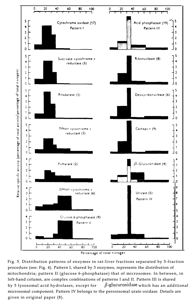
It is worth noting how remarkable this is. Popular conceptions of science often assume that new discoveries require directly "seeing" something new, like an image in a microscope. But De Duve, by developing new sample preparation and fractionation protocols for the ultracentrifuge and through careful analysis of enzyme activity across fractions, was able to infer the existence of an entirely new organelle — solely from patterns in his data. The combination of a new instrument with the tenacity of an experimentalist willing to develop new methods to creatively extract understanding is the hallmark of how new science instruments drive discovery.
Electron Microscope
…innovations in experimental technique can also radically alter the scene of inquiry in a science. New techniques make new forms of evidence available, and thus can lead to redefinition of standards of evidence and explanation in already established problem domains.
Picture Control, page 11
While the ultracentrifuge sprung from colloidal chemistry, new methods of seeing were often more explicitly borrowed from physics. The electron microscope was first built by Ernst Ruska in Berlin in 1933 and exploited the wave-like nature of electrons that allowed much higher atomic resolutions compared to optical microscopes, which are constrained by the wavelength of visible light.
The development of the electron microscope in the U.S. was accelerated by Radio Corporation of America’s (RCA) early interest. RCA represented the heyday of American manufacturing and innovation, a pioneer manufacturer of radios, vacuum tubes, and phonographs. In the late 1930’s, RCA embarked on an enormously expensive scientific prestige project to build an electron microscope, in part to rebuff claims from their competitors that their radio transmission technologies were inadequate.
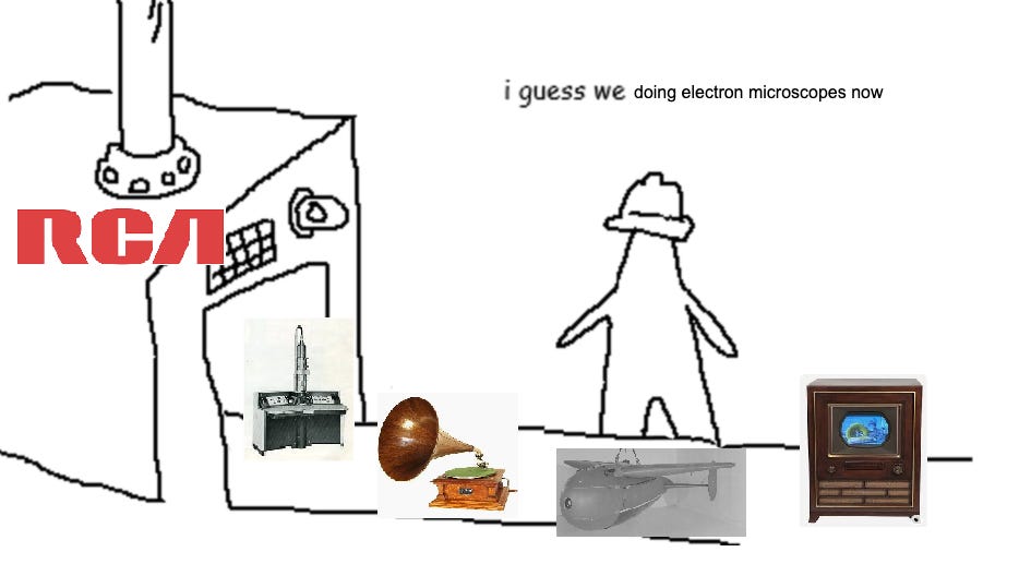
RCA’s first mover advantage lay in the simple fact that competitors like GE and Kodak correctly assessed that the market for the still nascent electron microscope was too small at the time. With the outbreak of World War Two, resources were no longer allocated towards making televisions, but RCA’s second electron microscope prototype, the Model B, was granted top defense priority (AA1 status) by the War Production Board, as a significant unclassified high-technology development to counter claims of German scientific superiority.3 It helped that RCA had also funded a National Research Council (NRC) electron microscopy fellowship at their corporate R&D lab in Camden, New Jersey.4 Their electron microscope center helped gatekeep early access to electron microscopy and allow the gradual development of an accepted methodology for preparing and interpreting electron micrographs.
While RCA’s NRC fellowship helped build an early community around their electron microscope, Weaver also situated the electron microscope at the center of his plans to revive the flailing biology department of MIT. With MIT president Karl Compton, Weaver brought in Francis Schmitt, a physiologist Weaver had cultivated while he was a faculty member at Washington University, to chair a new biology department at MIT. Schmitt’s arrival at MIT was bolstered by a $70,000 grant (~$1.5M in 2025 terms) from Weaver for “development and research centering around the new precision tool, the electron microscope”, including the procurement of a RCA Model B electron microscope. Schmitt would go on to pioneer the field of biophysics, with a department known for its doubly rigorous biology and physics training curriculum for graduate students. While Schmitt never won a Nobel Prize, he was instrumental in developing foundational techniques for operating the electron microscope and produced some of the first electron micrographs of collagen, myelin sheaths, and nerve fibers.
In parallel to Schmitt’s work at MIT, Albert Claude and Keith Porter at Rockefeller Institute (institutionally separate from Rockefeller Foundation, but both well known to Weaver), were also experimenting with electron microscopes.5 They produced the first electron micrographs of intact cells in the mid-1940s, revealing the endoplasmic reticulum — a vast, folded membrane system that no one had known existed. George Palade, who joined Rockefeller in 1947, then used the electron microscope to discover ribosomes studding the endoplasmic reticulum and trace the pathway by which proteins are secreted from cells. Claude and Palade would share the 1974 Nobel Prize with De Duve, whose ultracentrifuge work we covered above, for their discoveries of cell organelles. Together, they transformed the cell from what Claude called a mass “as distant from us as the stars and galaxies” into a system of known organelles with known functions — all made visible by the electron microscope and the ultracentrifuge working in tandem.
It is worth remarking how prescient of a conviction Weaver had in electron microscopes. From the outset, it was not obvious electron microscopes, with their vacuum sample chambers and intense ionizing beams, could be applied to biological samples, as opposed to the more inert inorganic metals they were a more natural fit. Several immediate challenges were apparent. First, biological samples would dehydrate rapidly under vacuum and beam conditions. Second, samples had to be made thin enough that electron detectors could measure deflection accurately on the backside. And finally, high contrast had to be induced, as the tiny electrons often had little deflection through water-filled biological samples. Addressing these problems required a stack of new techniques. Creating consensus around these methods took even longer. Debates over whether what electron microscopists were seeing was real biology or preparation artifact consumed the field for over a decade, with traditional biologists often pushing back to defend their own turf.
Ultimately, the electron microscope brought tremendous perceptive precision, moving us past the wavelength barrier of optical microscopes. Weaver’s early conviction to encourage the application of the electron microscope towards biology accelerated the development of new techniques and methods to make the electron microscope hospitable for viewing cells. Today, the electron microscope remains a workhorse of materials and biology characterization, particularly with the new revolution in cryoEM6.
Even now, we can still see the echoes of Weaver’s concentrated grantmaking around the electron microscope. In 1949, Weaver made a large grant to Cornell to establish an all-university Electron Microscope Center, notably under the engineering physics department, rather than a biology department. Today, Cornell is still a leader in electron microscopy techniques, with two Cornell researchers developing a new electron detector (the EMPAD), which was licensed to Thermo Fisher and is now ubiquitous in high-end electron microscopes.
X-Ray Crystallography
…making use of recent developments in physics. X-ray diffraction by crystals and electron diffraction by gas molecules have been the experimental methods principally used. The theoretical work involves the application of quantum mechanics to complex molecules. The work has now advanced to a point where it becomes possible to study the structure of chlorophyl, hemoglobin, and other substances of basic biological importance…
Rockefeller Foundation Annual Report, 1933
Like the electron microscope, X-ray crystallography was first used by physicists for studying crystals and minerals. Following von Laue winning the 1914 Nobel Prize for discovering crystal X-ray diffraction, the father-son Bragg duo won the 1915 Nobel Prize for developing a quantitative understanding of how to interpret X-ray diffraction to study crystal structures.
When Weaver joined Rockefeller foundation, one of the first grants he made was to fund the application of X-ray crystallography, previously used for inorganic crystals, to squishy biological compounds. In 1932, he supported the famous Linus Pauling’s lab at Caltech, including for the development of “an x-ray method, by means of which the environment of an iron atom in hemin and related substances can be investigated.”
In 1936, Max Perutz, a chemistry graduate from Vienna, arrived at Cambridge’s Cavendish Laboratory to work with J.D. Bernal, who had recently demonstrated that wet protein crystals could produce sharp X-ray diffraction patterns. When Lawrence Bragg became Cavendish Professor in 1938, he saw promise in Perutz’s work on hemoglobin crystals and championed it as exactly the kind of problem where X-ray methods could open up biology — a conviction he shared with Weaver. From 1939, the Rockefeller Foundation began funding Perutz and Bragg’s lab, sustaining their work with funding throughout the difficult war years in the U.K. and onwards.
Applying X-ray crystallography to proteins required solving two problems: crystallizing the protein, and then interpreting the resulting diffraction pattern. For minerals, diffraction patterns were clean and calculable based just on the unit cell of the mineral crystal. For a complex, folded protein with thousands of atoms, both steps were a nightmare. Perutz spent 15 years attacking the interpretation problem before cracking it in 1954 with isomorphous replacement — attaching heavy mercury atoms to the protein and comparing diffraction patterns with and without them. John Kendrew, a former military advisor to Mountbatten who had joined Perutz at the Cavendish in 1945, applied this method to the simpler protein myoglobin, producing the first 3D protein structure in 1958 with sperm whale myoglobin, after unsuccessfully trying to crystallize myoglobine from a half dozen mammals. Calculating protein structure from the resulting diffraction pattern was only possible because Kendrew invested the time to learn how to program Cambridge’s EDSAC, one of the world’s first electronic computers — yet another case of a discovery only being possible thanks to a new tool.7
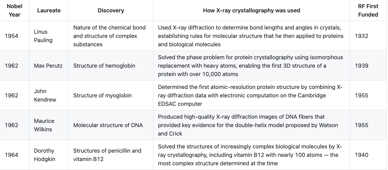
From the perspective of Nobel Prizes, X-ray crystallography was, by a significant margin, the single most productive instrument in Weaver’s portfolio. Pauling at Caltech, funded from 1932, used X-ray diffraction to establish the rules of chemical bonding, discover the alpha helix structure of proteins, and identify the molecular basis of sickle cell anemia — the first disease understood at the molecular level. Perutz and Kendrew at Cambridge, funded from 1939, spent over two decades developing the methods to solve the first protein structures, along the way training Francis Crick and hosting James Watson, who used the lab’s X-ray expertise to build their model of DNA. Maurice Wilkins and Rosalind Franklin at King’s College London used Rockefeller-funded X-ray equipment to produce diffraction images of DNA fibers that were the key experimental evidence for the double helix. Dorothy Hodgkin at Oxford, who had trained under Bernal at Cambridge and co-produced the first X-ray photographs of a protein crystal, used Rockefeller grants and an early IBM machine to solve the structure of penicillin during the war and then vitamin B12, cholesterol, and eventually insulin over a 35-year effort. 89
X-ray crystallography was so productive for the same reason any new instrument is productive for early adopters — the methodological knowledge required to make it work compounds over time and is very hard to transfer. Weaver, in his autobiography Scene of Change, includes a quote from Bragg describing this directly:
I received a letter from Sir W. Lawrence Bragg, the younger of the father-and-son team that received the Nobel Prize in 1915 for their determinations of crystal structures by X-ray diffraction techniques. In this letter Sir Lawrence said, “concerning the part the Rockefeller Foundation played in helping the ‘Cambridge School.’ Your help came at a vital time just before the war when I was trying to find some way of supporting Perutz’s work [Max F. Perutz was the chairman of the Laboratory of Molecular Biology at Cambridge] and it was continued after it. ‘This school was responsible for DNA, for the first protein structures, for the first understanding of virus structure, and for work on muscle.
‘The extent to which the X-ray analysis of protein was pioneer work is shown by the fact that only now, twelve years after the trail was beaten at Cambridge and the Royal Institution, has any other research centre succeeded in getting a protein ‘out.’ I am allowing myself to put this so strongly just because I think that the Foundation’s help made an outstanding difference to these advances.”
This is the paradox and promise of new science instruments: they are slow to yield results, but those results, once obtained, are so definitive and information-rich that they reshape entire fields. Weaver’s willingness to fund this kind of long-horizon, high-risk science instrumentation work — for nearly two decades before it produced a single solved protein structure — is perhaps the strongest evidence for the value of patient, conviction-driven science philanthropy, and the impact of a science-instrumentation driven view of scientific field creation.10
What can we learn from this?
Warren Weaver's tenure at the Rockefeller Foundation is held up as an exemplar of what a high-conviction program director can accomplish: the creation of a new scientific field, an explosion of research that directly benefits mankind, and a bevy of Nobel Prizes to boot.
A key ingredient to Weaver's success is not just funding physicists to work in biology, but bringing the tools of physics to the problem of biology. Through the application of new ways of seeing, like the electron microscope and x-ray crystallography, and new ways of separating, like the ultracentrifuge, Weaver-funded scientists were able to make novel discoveries with breakthrough hit rates. While only covered on the fringes in this post, Weaver’s instrument-driven thesis was orchestrated as part of a broader institutional and industrial strategy to create centers of excellence and drive commercial production of new tools.
The counterfactual impact of Weaver's funding lay in the high risk that comes with adopting new tools. For a scientist to introduce a new instrument, a new way of seeing the world, does not mean default acceptance of new methods by their peers. At the outset, scientists don't know whether a new instrument will yield new insights or be unworkable with the specimens and samples of interest. The ultracentrifuge was a difficult machine to engineer and new ramp rate protocols, suspension mediums specific to cells, and cell homogenization techniques were needed; electron microscopes were death rays for cells in a vacuum, and required new thin-sectioning, phase contrast media, and fixation protocols to prevent distortion of cell shapes; while interpreting X-ray diffraction patterns of proteins required the invention of the electronic computer and creative techniques to crystallize proteins and extract structural insight from diffraction patterns.
Weaver's funding and support for scientists willing to take a leap with him and commit their careers to a new science instrument — often because they came from a different domain and saw possibilities that biologists couldn't — is part of why his funding strategy was so impactful. His playbook for using new instruments to create new fields of science is still instructive today. In subsequent posts, I will write about my own "Weaver thesis", how to think about the role of AI as a new tool for scientists in different domains, and implications for U.S. science policy.
Total funding for Natural Sciences Experimental Biology during Weaver’s tenure was ~$600M (in 2025 dollars), though Weaver would often appropriate Rockefeller Foundation fellowship and aid grants for his purposes as well.
Claude and Palade would also win the Nobel Prize with De Duve, for discovering other cell organelles with electron microscopes.
Ruska would go on to work with Siemens to commercialize an electron microscope right before World War Two.
My thesis is that the death of the corporate R&D lab is what has enabled the consolidation of the science instrument industry.
CryoEM is notable in that it is also primarily defined as a methodological innovation in sample preparation.
There is some literature on the role compute plays in accelerating scientific discoveries. See also my work on the role of supercomputers in advancing weather forecasting.
Dorothy Hodgkin are both illustrative examples in X-ray crystallography.
The underappreciated role of science instrumentation is also paralleled in the role of women in science. Often, it was female scientists, brought in during the war, who were delegated the task of operating science instruments and “computers” at universities, and uranium centrifuges at the Manhattan Project. They were often pioneers of new methods for operating science instruments that are still foundational today and supported important discoveries, but forgotten in the story of scientific progress. Rosalind Franklin’s story is now well known, but even Dorothy Hodgkin winning the Nobel Prize was not sufficient to overcome gender stereotypes of the time. Schmitt’s lab at MIT for electron microscopy also saw Marie Jakus who developed several important techniques to enable clear contrast in electron micrographs but was never able to benefit in their career from the enormous amounts of science their established methods enabled.
It is also an example of the importance of corporate R&D labs. Bell Labs Nobel Prize discovery of electron diffraction was made possible through building a new science instrument, which took 30 years to ultimately commercialize at scale.
But the Davisson and Germer set-up was so unique, it was actually the first pioneering example of low-energy electron diffraction (LEED), a new surface characterization technique Germer would go on to pioneer in the 1960’s. The 30 year gap between their experiment and widespread commercialization was due to inadequate detector quality and the lack of commercially available high vacuum pumps, demonstrating just how far ahead of their time this experimental setup was.
I enjoyed this piece and I think there’s plenty of truth to it. For something with a broader view of an enabling technology you might like The Glass Bathyscaphe by Alan Macfarlane and Gerry Martin. I do think it can be overstated though. The Rockefeller foundation spent heavily on Edward Lawrence’s cyclotrons in the 30s but it was Irene and Frederic Curie who invented the technique of bombardment with alpha particles to generate new elements. Their experimental setup was almost absurdly primitive. The instrument builders were unable to perceive that insight. Finally Bret Victor’s Seeing Spaces lecture is I think inherently related to the ideas here.
This piece was excellent and I’m very excited to read about your thesis in subsequent posts!
This question is out of scope, but I’m curious if and when the Rockefeller Foundation deviated from this approach of funding instruments (i.e. did this approach live and die with Weaver), and if they’ve had similar funding successes post 1950? Where did they go wrong, if not?
I’d also love to see you write about intuitions you have about underlying techniques that currently exist that need better tools to fully appreciate their outputs.
For example: “For the ultracentrifuge, cell homogenization, the ability to shred cell walls without damaging the organelles that were to be separated; appropriate suspension mediums, to prevent distortion of the organelles in solution; and centrifugation ramp rate protocols, to provide clean separation of organelles; all had to be tested and developed before biologists could even begin to use the ultracentrifuge to understand cellular components and structure.”
What is an equivalent of discovering how to shred cells walls, but not having an ultracentrifuge to understand the cellular components that exists today that you think is important to create?
Thanks for writing this :)